


Eye Anatomy and Major Diseases
Eye Anatomy and Major Diseases: How We See and What Matters
Our eyes are complex and remarkable organs that receive light and transmit it to the brain. Acting like sophisticated cameras, various parts work in harmony to allow us to see the world clearly. In this blog post, we'll explore the main structures of the eye and the common diseases associated with each part.
1. Outermost Layer of the Eyeball: Protection and Light Passage
- Sclera: This is the thick, tough, white outer layer that surrounds the eyeball. It maintains the eye's shape and protects its internal structures. What we commonly call the "white of the eye" is the sclera.
-
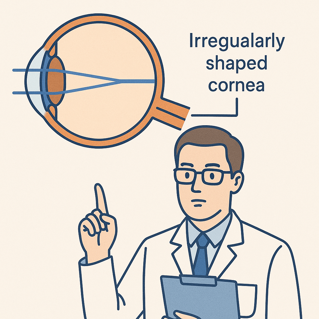
Cornea: A transparent membrane located at the front of the sclera, it is the first part to refract (bend) light entering the eye. It remains transparent because it has no blood vessels, and it's a crucial component for vision. LASIK and LASEK surgeries correct vision by altering the shape of this cornea.
-
Related Diseases:
- Keratitis: Inflammation of the cornea, causing pain, redness, and vision reduction. Improper contact lens use is a major contributing factor.
- Keratoconus: A condition where the cornea gradually thins and bulges into a cone shape, leading to severe astigmatism and vision loss.
-
Related Diseases:
-
Conjunctiva: A thin, transparent membrane that lines the inside of the eyelids and covers the white part of the eyeball. It helps protect the eye and distribute tears evenly.
-
Related Diseases:
- Conjunctivitis: Inflammation of the conjunctiva, often caused by bacteria, viruses, or allergens, resulting in redness, itching, and discharge (pink eye).
-
Related Diseases:
2. Middle Layer of the Eyeball: Light Regulation and Nutrient Supply
-
Uvea: This is the middle layer located inside the sclera, rich in blood vessels and pigments. It plays a vital role in supplying nutrients to the eye and regulating the amount of light entering it. The uvea is divided into three parts:
- Iris: The colored part of the eye that controls the size of the pupil to regulate the amount of light entering the eye. The pupil constricts in bright light and dilates in dim light.
- Ciliary Body: Crucial for supporting the lens and changing its shape to adjust focus. It also produces aqueous humor, the fluid that fills the front part of the eye.
- Choroid: Located beneath the retina, this highly vascular membrane supplies oxygen and nutrients to the retina.
-
Related Diseases:
- Uveitis: Inflammation of the uvea, which can cause vision loss, pain, redness, and light sensitivity. Severe cases can lead to blindness.
- Pupil: The central opening in the iris through which light enters the eye. Its size changes as the iris contracts or dilates.
-
Lens: A transparent, biconvex structure located behind the pupil. Its thickness is adjusted by the ciliary body to focus light precisely on the retina, much like a camera lens.
-
Related Diseases:
- Cataract: A condition where the lens becomes cloudy, preventing light from passing through properly. Vision appears foggy or hazy, and glare may be significant in bright light. Primarily caused by aging.
-
Related Diseases:
3. Innermost Layer of the Eyeball: Light Detection and Signal Transmission
-
Retina: The innermost layer of nerve tissue in the eye, comparable to a camera's film. It contains photoreceptor cells (rods and cones) that convert light into visual signals. Rods detect light and dark in dim conditions, while cones perceive color in bright conditions.
-
Related Diseases:
- Retinal Detachment: A severe condition where the retina separates from the underlying supportive tissue of the eye. Without prompt treatment, it can lead to permanent vision loss. Symptoms include floaters, flashes of light, and a shadow in the peripheral vision.
- Diabetic Retinopathy: A complication of diabetes where the blood vessels in the retina are damaged, leading to bleeding, swelling, and vision loss.
-
Related Diseases:
-
Macula: The central part of the retina with the highest concentration of photoreceptor cells, responsible for sharp, detailed central vision. We use the macula for activities like reading and driving.
-
Related Diseases:
- Macular Degeneration: Damage to the macula that causes progressive vision loss or distortion, particularly affecting central vision. Age-related macular degeneration (AMD) is the most common form.
-
Related Diseases:
-
Optic Nerve: A bundle of nerve fibers that transmits visual signals from the retina to the brain. Damage to the optic nerve can result in visual field defects or vision loss.
-
Related Diseases:
- Glaucoma: A disease characterized by damage to the optic nerve, often due to elevated intraocular pressure, leading to progressive visual field loss and potential blindness. Early detection through regular check-ups is crucial as initial symptoms are often absent.
-
Related Diseases:
-
Vitreous Humor: A clear, jelly-like substance that fills the space between the lens and the retina. It helps maintain the eye's shape and provides nutrients to the retina.
-
Related Diseases:
- Vitreous Floaters: Caused by tiny clumps or strands within the vitreous that cast shadows on the retina, appearing as small dots or strings floating in the field of vision. While mostly a natural aging process, a sudden increase or change can indicate a more serious retinal issue.
-
Related Diseases:
As demonstrated, each part of the eye is intricately connected, working together to convert light into electrical signals and transmit these signals to the brain, allowing us to 'see' the world. Understanding that the health of each component directly impacts our vision is crucial.
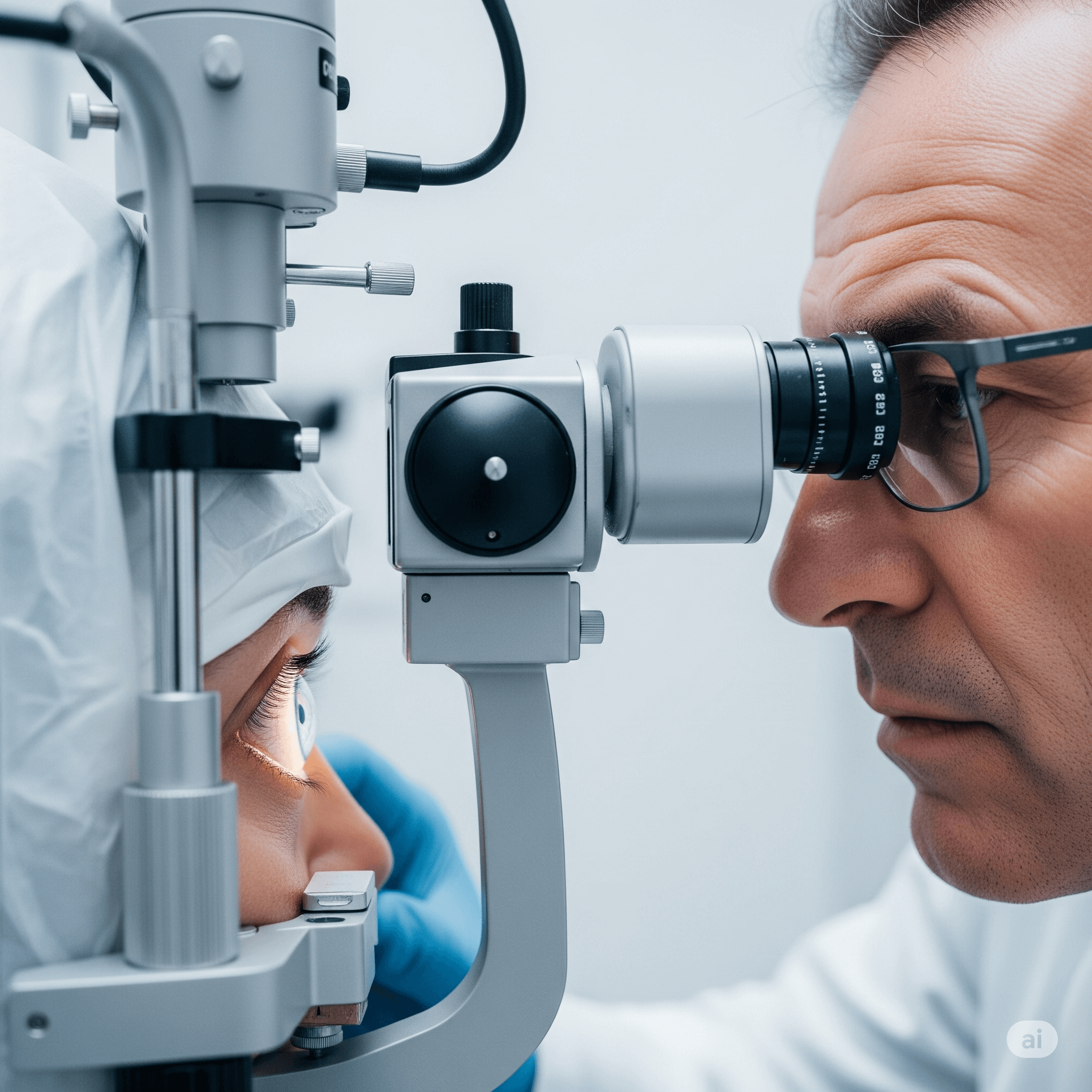
If you experience any discomfort in your eyes or notice changes in your vision, do not hesitate to visit an ophthalmologist for a professional diagnosis and consultation!
Related Posts
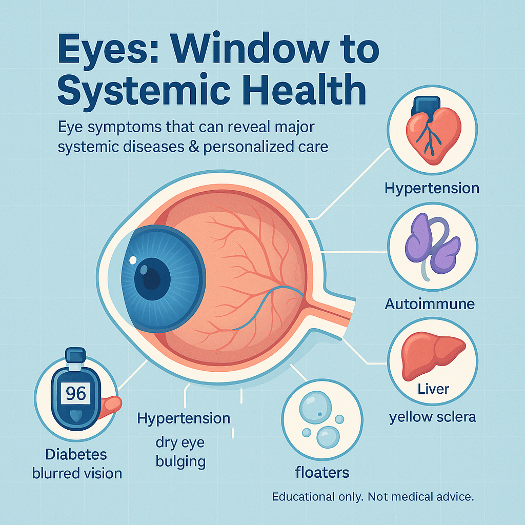
Your Eyes, Your Health: The Unexpected Connection

🥦 Eye Health Guide: Science-Backed Diet and Exercise Tips for Better Vision
Genetics and Vision: How Your DNA Shapes Eye Health
Why Astigmatism-Correcting Color Contact Lenses Are Essential | LUMINIX Toric Lenses
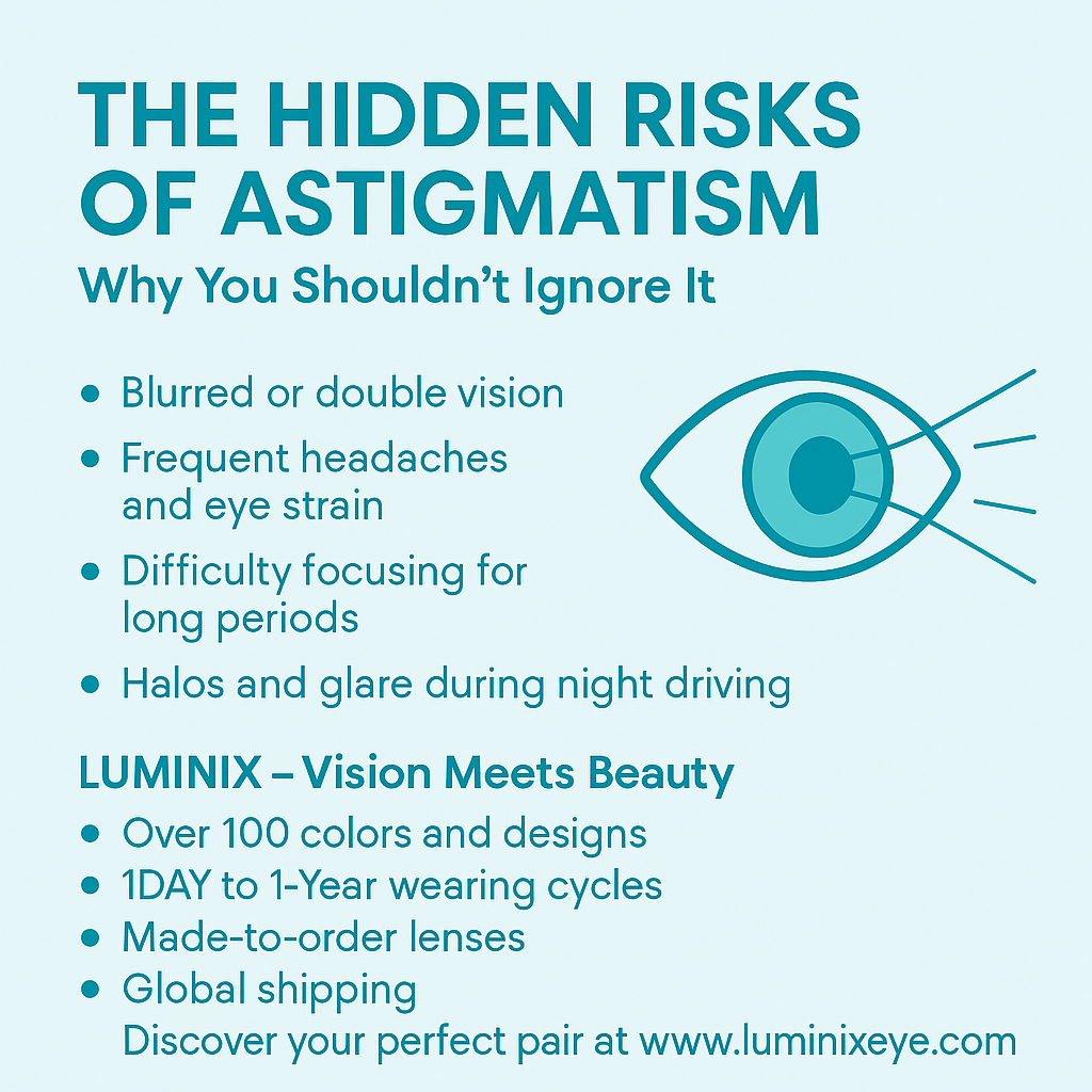
Astigmatism Risks and Solutions | LUMINIX Toric Color Contact Lenses

TORICA: More Than Just Colored Lenses